Nasopharyngeal cancer staging – 5 minute review

In this video, Dr. Katie Bailey takes us quickly through the nasopharynx, a common location of malignancies in the head and neck, and walks us through some quick samples of how they are staged.

Review of the anatomy of the nasopharynx. The nasopharynx is behind the palate and includes the clivus.  The nasopharynx is largely lined by squamous mucosa with lymphoid tissues and muscles. An important landmark is the torus tubarius and fossa of Rosenmuller.   

Nasopharyngeal cancer staging, like other subsites, is based on a T, N, M stage. Nasopharyngeal cancers are predominantly staged based on whether adjacent structures, such as surrounding soft tissues or the skull base, are involved. Unlike the other tumor sites, size is not involved in the T staging.

Example case 1. There is a soft tissue mass filling the nasopharynx, asymmetrically larger on the right. Erosion of the adjacent bony structures, including the clivus, are important. The tumor is extending into the sphenoid sinus. Involvement of the bony structures and sinuses makes this is a T3 tumor, and there were no nodes or distant metastases (not shown).

There is a more subtle example of bone erosion of the posterior wall of the sphenoid sinus and left carotid canal along with the clivus.

Example case 2. There is a soft tissue mass eroding the petrous apex and left aspect of the clivus. MRI better shows the extent of the involvement, where you can see that there is intracranial involvement into Meckel’s cave (trigeminal foramen). The intracranial involvement makes this a T4 tumor. There were no nodes or distant metastases (not shown).

Example case 3. In this case, there are necrotic lymph nodes in the parapharyngeal and prevertebral space as well as extending down the neck at multiple internal jugular levels. The original CT did not show a mass in the nasopharynx, but because of the nodes, a nasopharyngeal cancer is suspected. The PET/CT is able to locate the abnormality along the left torus tubarius. The local nature of this tumor makes it T1, but the bilateral lymph nodes make it an N3 for nodes.

Extra case. An incidental polypoid mass is seen in the left nasopharynx on a brain MRI with minimal peripheral enhancement and central reduced diffusion. The homogeneously low T2 appearance and reduced diffusion make this suspicious for lymphoma, although you would not be able to tell until this lesion had been biopsied.

Thanks for checking out this quick video on nasopharyngeal cancer staging. Be sure to tune back in for additional videos on staging of the other head and neck subsites.Also take a look at the head and neck topic page as well as all the head and neck videos on the site.


See this and other videos on our Youtube channel

Oropharyngeal cancer staging – 5 minute review

revised 11/12/2022
N.B. The initial version of this video had a few small errors in the staging, so we have corrected them.

In this video, Dr. Katie Bailey describes the anatomic subsites of the oropharynx and reviews how tumors are staged through four quick example cases.

Review of oropharynx anatomy. The oropharynx includes the tonsils (both lingual and palatine), the squamous mucosa of the pharynx, the uvula, and the vallecula.   oral cavity includes the lips, teeth, hard and soft palate, gingiva, retromolar trigone, the buccal mucosa, and anterior 2/3 of the tongue. The masticator space contains the muscles of mastication, the mandible, branches of the trigeminal nerve, lymph nodes, and minor salivary glands. 

Oropharyngeal cancer staging. Tumor (T) staging is based on the size of the tumor or invasion through adjacent structures. Nodal (N) staging is based on the number, location, and size of nodes, and metastasis (M) staging is based on the presence or absence of distant sites of disease.

Example case 1. There is a 2.4 cm mass of the right palatine tonsil. There is level 2 and 3 adenopathy. The lymphadenopathy compresses the jugular vein and displaces the adjacent sternocleidomastoid.  The size of the tumor makes this a T2 lesion, and the unilateral adenopathy less than 6 cm with multiple nodes makes it N2b. Because metastases can’t be evaluated with this information, it is given an ‘X’ for M staging right now.

Example case 2. There is a 3.2 cm mass in the tongue base and extending into the vallecula. There is no extension into the adjacent structures or fat. There is a single left sided level 2 lymph node that is somewhat prominent but isn’t definitely abnormal. That makes this a T2N0Mx tumor. If a PET or biopsy later shows that the node is positive, the staging can be changed.

Example case 3. There is a subtle mass of the right lateral wall of the oropharynx involving the tonsillar pillar and tongue base. This one is quite hard to see. There are cystic necrotic lymph nodes on the right, but none greater than 6 cm. A PET/CT showed no distant metastatic disease. That makes this a T1N2bM0 tumor.

Example case 4. This patient presented with cervical lymphadenopathy on the left but had no clear primary tumor in the oropharynx. There was no mass of the tongue base or elsewhere. The patient had a lung node suspicious for metastatic disease. A PET/CT showed that there was a primary in the soft palate. The mass was detected only by PET/CT. The final staging for this cancer is T1N2bM1.

Thanks for checking out this quick video on oropharyngeal cancer staging.

Thanks for checking out this quick video on oral cavity cancer staging. Be sure to tune back in for additional videos on staging of the other head and neck subsites. Also take a look at the head and neck topic page as well as all the head and neck videos on the site.


See this and other videos on our Youtube channel
Oral cavity cancer staging – 5 minute review

In this video, Dr. Katie Bailey takes on a common topic and describes how cancers of the oral cavity are staged through three quick example cases.

Review of oral cavity anatomy. The oral cavity includes the lips, teeth, hard and soft palate, gingiva, retromolar trigone, the buccal mucosa, and anterior 2/3 of the tongue.

Retromolar trigone. The retromolar trigone is a common crossroads where a lot of pathology occurs and it can cause referred ear pain. Mandibular invasion can also occur with masses that occur in this location.

Oral cavity cancer staging. Tumor (T) staging is based on the size of the tumor and depth of invasion. Nodal (N) staging is based on the number, location, and size of nodes, and metastasis (M) staging is based on the presence or absence of distant sites of disease.

Example case 1. This example case involves the oral tongue and measures 2-4 cm. It has ipsilateral lymph nodes less than 3 cm. This makes this a T2N2 tumor. Because metastases can’t be evaluated with this information, it is given an ‘X’ for M staging right now.

Example case 2. This shows a subtle left tongue cancer measuring less than 2 cm. There are no lymph nodes. That makes this a T1N0Mx tumor.

Example case 3. This case is a hard palate tumor with bone erosion. The involvement through adjacent structures (the bone of the hard palate) makes this a T4 tumor. There is no nodal involvement, making this a T4N0Mx cancer.

Thanks for checking out this quick video on oral cavity cancer staging. Be sure to check out the head and neck topic page as well as all the head and neck videos on the site.

See this and other videos on our Youtube channel

Neuroradiology Board Review – Brain Tumors – Case 20 – Summary

Neuroradiology brain tumor board review. This lecture is geared towards the ABR core exam for residents, but it would be useful for review for the ABR certifying exam or certificate of added qualification (CAQ) exam for neuroradiology.

This video has both the final case of the series and a quick review summary!

Case 20

In this case, you are starting with an immunocompromised patient with HIV. Initial CT images show a hyperdense mass in the left basal ganglia with a lot of surrounding edema. This is helpful, because a few things are known for being hyperdense on CT.

MRI images confirm a mass in the basal ganglia. It is somewhat T2 hypointense with well-defined margins and surrounding edema. On postcontrast images, it has peripheral enhancement but central non-enhancement compatible with necrosis.

The differential diagnosis for a solitary enhancing parenchymal mass is different in an immunocompromised patient (or someone on immune suppressing agents. In an immune normal patient, the top diagnoses are

  • metastatic disease
  • high grade glioma
  • lymphoma

On the other hand in an immunocompromised patient the order of these diagnoses shifts to include:

  • lymphoma
  • infection
  • metastatic disease
  • high grade glioma

As you can see, lymphoma and infection jump to the top in an immunocompromised patient.

The diagnosis is: CNS lymphoma

CNS lymphoma can occur when associated with systemic lymphoma or primarily in the CNS, as in this case. This is most commonly a diffuse large B-cell lymphoma. It is more common in immune compromised patients. It often occurs in the basal ganglia and periventricular white matter and can often be multifocal. Lymphoma is one of the rare diseases which is T2 hypointense, so you should think about it if you see a T2 hypointense mass.

In immunocompetent patients, lymphoma most commonly has solid enhancement. However, in immunocompromised patients it is much more likely to show central necrosis, as in this case. Also, in an immunocompromised patient, it can be hard to differentiate lymphoma from infection, particularly toxoplasmosis. The two most common ways to try to differentiate this are to start a trial of toxoplasmosis therapy for a few weeks and see if the lesions improve and to perform a thallium-201 chloride nuclear medicine scan. Lymphoma has thallium uptake, while toxoplasmosis does not.

Summary

In this board review lecture, you’ve seen a lot of different tumors and how they manifest in different situations. In many cases, you can’t make a definitive diagnosis but you should always be able to come up with a reasonable differential diagnosis. It’s also helpful to know some of the basics about treatment and prognostic factors.

There are two key strategies that I hope can help you get a few additional points, the approach to CP angle masses and the approach to cortical tumors.

Cerebellopontine angle masses

As we’ve seen in some of the other cases, cerebellopontine angle masses can be solid or cystic. Solid masses that involve the IAC and expand it are likely schwannomas, while others outside the IAC are likely meningiomas. Arachnoid cysts and epidermoids are the most common cystic masses which are differentiated by DWI (which is bright in ependymomas.

Cortical tumors

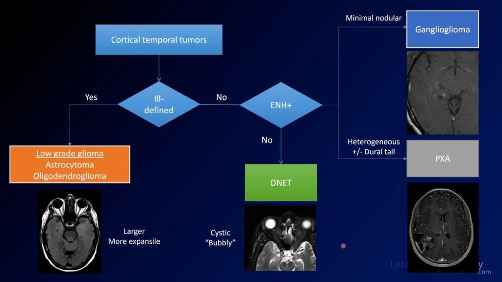
Several of the cases in this series dealt with cortical temporal tumors. Ill-defined masses that are larger are more likely to be low grade gliomas (oligodendrogliomas and astrocytomas). Completely non-enhancing bubbly masses favor DNET. A little nodular enhancement favors ganglioglioma, while pleomorphic xanthoastrocytoma (PXA) can be more avidly enhancing and irregular.

Neuroradiology Board Review – Brain Tumors – Case 19

Neuroradiology brain tumor board review. This lecture is geared towards the ABR core exam for residents, but it would be useful for review for the ABR certifying exam or certificate of added qualification (CAQ) exam for neuroradiology.

More description and the answer (spoiler!) are seen below the video.

This case of a patient with diplopia (double vision) starts with a CT through the orbits. You can see there is a well-defined mass at the right orbital apex.

On MRI, you can see the lesion again is well-defined with well defined margins and is hyperintense on T2. On T1, the lesion is isointense to muscle on pre-contrast and then demonstrates heterogeneous enhancement on postcontrast. It looks like the lesion is enhancing more on the coronal image compared to the axial image.

The diagnosis is: orbital venous vascular malformation

Orbital venous vascular malformations are sometimes referred to as hemangiomas, although this term is falling out of favor because it is not neoplastic (unlike the neoplastic infantile and neonatal orbital hemangiomas). These are relatively benign lesions that can cause visual problems secondary to mass effect, but it’s relatively uncommon for them to enlarge.

One characteristic finding of orbital venous malformations is progressive enhancement on delayed images. On early images, it might be enhancing a little bit but if you have more delayed images, you might see more enhancement. If you don’t have more than one plane of contrast, you can bring them back and image them again 15-30 minutes later and it should fill in.

The primary differential considerations for orbital masses are sarcoidosis, idiopathic or IgG4 related orbital disease, metastatic disease, and lymphoma. When it is this well defined and has the characteristic delayed enhancement, the diagnosis is relatively certain.

Neuroradiology Board Review – Brain Tumors – Case 18

Neuroradiology brain tumor board review. This lecture is geared towards the ABR core exam for residents, but it would be useful for review for the ABR certifying exam or certificate of added qualification (CAQ) exam for neuroradiology.

More description and the answer (spoiler!) are seen below the video.

This case starts with 2 axial images from a CT through the posterior fossa followed by MRI images through the same region. There is a heterogeneous lesion to the left of the midline along the left foramen of Lushka. On postcontrast images, it is pretty avidly enhancing. The enhancing margins are pretty well defined and it looks like it is wholly in the ventricle.

The diagnosis is: ependymoma

Ependymomas are enhancing intraventricular tumors arising from the ependymal lining. They are commonly enhancing and conform to the ventricles and the ventricular outflow tract, which results in their description of “toothpaste” like lesions. In adults, they most commonly occur in the 4th ventricle although in pediatric patients they can occur elsewhere.

When in the posterior fossa, your main differential is choroid plexus papilloma/tumor. If you can’t tell that it’s an intraventricular lesion it can be harder because the differential diagnosis also include metastatic disease and possibly medulloblastoma. If you see a lesion that looks similar but doesn’t enhance very much, think about it’s sister lesion subependymoma.

Neuroradiology Board Review – Brain Tumors – Case 17

Neuroradiology brain tumor board review. This lecture is geared towards the ABR core exam for residents, but it would be useful for review for the ABR certifying exam or certificate of added qualification (CAQ) exam for neuroradiology.

More description and the answer (spoiler!) are seen below the video.

In this case, we have an MRI showing a FLAIR and T2 hyperintense mass in the left insula with relatively ill-defined margins. On SWI, there are some areas of susceptibility that probably represent calcification, although blood products could look similar. Postcontrast images demonstrate little or no contrast enhancement.

1:33 The diagnosis is: oligodendroglioma

Oligodendrogliomas are gliomas which are now defined by the characteristic genetic features of IDH mutation and 1p19q codeletion (loss of portions of both chromosomes 1 and 19). They can be WHO grade 2 (as in this case) or grade 3 (anaplastic oligodendroglioma). Theoretically, these lesions never degrade into WHO grade 4 lesions although the grade 3 lesions can be quite aggressive. In general, oligodendrogliomas have a better prognosis than their sister gliomas, astrocytomas. They respond better to radiation and have better overall survival.

Oligodendrogliomas are treated with a combination of resection and chemoradiotherapy.

The susceptibility seen within the tumor on this case represents areas of calcification. Oligodendrogliomas are one of the main considerations if you see an expansile tumor with calcification.


Neuroradiology Board Review – Brain Tumors – Case 16

Neuroradiology brain tumor board review. This lecture is geared towards the ABR core exam for residents, but it would be useful for review for the ABR certifying exam or certificate of added qualification (CAQ) exam for neuroradiology.

More description and the answer (spoiler!) are seen below the video.

This case shows a 20 year-old with seizures. MRI shows a lesion in the medial temporal lobe along the tentorium. It is relatively well circumscribed with a cystic portion as well as an enhancing nodule. There is not much mass effect.

The diagnosis is: ganglioglioma

Gangliogliomas are low grade tumors often found in the temporal lobes of patients with seizures. They are mixed in their cell origin, containing both components of glial and neuronal cells. Along with pilocytic astroctyoma, PXA, and hemangioblastoma, they are one of the tumors in the differential for a tumor with a cyst and a nodule. They can be impossible to differentiate from DNET, but if you see enhancement it is more likely to be a ganglioglioma. Gangliogliomas are the most common neoplastic cause of epilepsy.

When you see a minimally enhancing cortical tumor, you should have a relatively short differential which includes:

First consider whether they are ill-defined or well-marginated. If ill-defined, the differential includes astrocytoma or oligodendroglioma. If well-marginated, then consider whether there is enhancement. If no enhancement, DNET is most likely. If there is a small amount of nodular enhancement, favor ganglioglioma, as in this case.


In a testing situation, if a small and minimally enhancing cortical tumor enhances a little bit, choose ganglioglioma. If you don’t seen enhancement, choose DNET. PXAs tend to be much more heterogeneous and irregular.

If you use a structured approach to these tumors, you can fall back on it if you aren’t really sure what you are looking at. With these relatively simple rules, you can be sure to get the most points on your exams AND give the most meaningful differential diagnosis.

Full Brain Tumor Board Review Playlist on Youtube

Neuroradiology Board Review – Brain Tumors – Case 15

Neuroradiology brain tumor board review. This lecture is geared towards the ABR core exam for residents, but it would be useful for review for the ABR certifying exam or certificate of added qualification (CAQ) exam for neuroradiology.

More description and the answer (spoiler!) are seen below the video.

As we start this case, we see a brain and a bone window from a CT through the posterior fossa. It is a little bit hard to see because it is a midline abnormality. The sagittal and coronal reformats are helpful in identifying where the lesion is located.

MRI is a little bit easier to see the lesion, but only a little. There is a well-demarcated lesion along the inferior aspect of the 4th ventricle. There is calfication, which you could see both on CT and GRE sequences from MRI. On post-contrast imaging, there is little, if any enhancement.

The diagnosis is: subependymoma

Subependymomas are relatively benign tumors arising from the walls of the ventricles. In contrast to most other intraventricular tumors, they often do not have much enhancement. The most common locations are in the 4th ventricle and lateral ventricles. Many times they are incidental but they can cause symptoms related to hydrocephalus.

Your differential diagnosis for intraventricular lesions includes meningioma, ependymoma (more enhancing), choroid plexus tumors (also more enhancing), and other tumors such as subependymal giant cell tumors (SEGT, often seen in patients with tuberous sclerosi).

Some tumor types have characteristic histologies that you should be familiar with for the test. Ependymomas are associated with perivascular pseudorosettes, while other tumors such as glioblastoma have their own histologic keywords (pseudopallisading necrosis, microvascular proliferation). These are pretty low yield to study but if you see them it’s good to be familiar with them.

Neuroradiology Board Review – Brain Tumors – Case 14

Neuroradiology brain tumor board review. This lecture is geared towards the ABR core exam for residents, but it would be useful for review for the ABR certifying exam or certificate of added qualification (CAQ) exam for neuroradiology.

More description and the answer (spoiler!) are seen below the video.

This case shows an MRI with a mass in the left parietal region near the midline. It is pretty well circumscribed and is probably extra-axial. It is homogeneously enhancing on postcontrast, and both T2 and T1 images demonstrate significant vascular flow voids suggesting that this is a very vascular lesion.

The diagnosis is: solitary fibrous tumor

Solitary fibrous tumors of the CNS (previously known as hemangiopericytomas) are similar in histology to fibrous tumors elsewhere in the body, such as the pleura. These were originally thought to be related to meningiomas, but their genetics has proven this to be false. However, the imaging features are similar to aggressive meningiomas. They are most often extra-axial, have avid enhancement, and broad dural attachments. It is common to have flow voids showing a high degree of vascularity.

Like meningiomas, these can be highly vascular lesions with much of their blood supply coming from external carotid branches. This angiogram shows the high degree of vascularity. It can be favorable to embolize as many of these vessels as possible prior to surgical resection to minimize bleeding.

Remember that the nomenclature has changed but you still may run into the term hemangiopericytoma. It’s something that should be on your differential if you see something that looks like an aggressive meningioma.