Vascular capstone course
Welcome to the Emory vascular capstone course. In this course, we are going to learn about vascular imaging of the head and neck. We’ll start with some general principles, such as the types of imaging that are used and when you might want to perform them. Then, you’ll have some interactive cases you can use to follow along and learn how to read vascular imaging on your own.
The following links will help you get access to the cases you need to scroll through the course and give you some additional information.
Before you start with the cases on your own, watch this video to learn some general principles of vascular imaging, including what types of studies we normally do and when you might want to do them.
Normal case
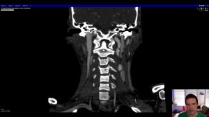
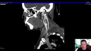
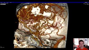
This is a case of a 30-year-old woman with a cervical spine fracture after trauma. Because of the extent of injuries, she underwent a CT angiogram to assess her vascular structures. In this case, the vasculature of her neck is normal. Use this case to practice your search through the vessels of the head and neck for injury.
Images
Top left – axial images from a CT angiogram
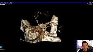
Top right – 3D volume rendering of the circle of Willis
Bottom left – oblique images through the right carotid artery
Bottom right – oblique images through the left carotid artery

Now that you know general concepts about imaging of the head and neck, you should learn about different types of pathology that you might encounter. Aneurysms, vascular malformations, dissections, and other abnormalities can all be seen on vascular imaging. Watch the following video before you proceed to the cases for independent learning.
Case A
80 year-old woman with altered mental status
Images
Top left – axial images from a CT head
Top right – 3D volume rendering of the circle of Willis
Bottom left -axial images from a CT angiogram
Questions
- On the non-contrast head CT (top left panel), what is the bright material and where is it located?
- What is the appropriate recommendation for the next imaging test after noncontrast CT?
- What are the abnormalities on CTA? Which vessels are affected?
Video explanation – Case A
Before you move on to the next case, check out the video explanation here.
Case B
47 year-old woman with new neurologic symptoms
Images
Top left – diffusion weighted images from an MRI.
Top right – axial images from a CT angiogram
Bottom left – oblique images through the right carotid artery
Bottom right – oblique images through the left carotid artery
Questions
- Where is the vascular abnormality on the CTA?
- What is the abnormality?
- What are the risks/complications if this is not treated?
Video explanation – Case B
Before you move on to the next case, check out the video explanation here.
Case C
43 year old woman with seizure
Images
Top left – axial images from a CT head
Top right – 3D volume rendering of the circle of Willis
Bottom left – axial images from a CT angiogram
Bottom right – coronal images from a CT angiogram
Questions
- What is the bright stuff on the noncontrast CT?
- What is the appropriate recommendation for the next imaging test after CT?
- What is the abnormality on CTA? What is the major risk if this is not treated?
- Bonus: What’s the next exam you should recommend?
Video explanation – Case C
Before you move on to the next case, check out the video explanation here.
Case D
41 year-old man after trauma from motor vehicle collision
Images
Top left – axial images from CT angiogram
Bottom left – coronal images from CT angiogram
Right – 3D volume rendering of left carotid artery
Questions
- Where is the abnormality located?
- What is the likely cause of the abnormality?
- Is this abnormality of the left internal carotid considered a “true” or “pseudo” version?
Video explanation – Case D
Before you move on to the next case, check out the video explanation here.