Brain imaging capstone course
Patients often present in the clinic and hospital settings with neurologic symptoms including headache, dizziness, seizure, weakness, and numbness. For this reason, brain imaging with either computed tomography (CT) or magnetic resonance imaging (MRI) is one of the most common radiology exams, and every physician is likely to encounter imaging of the brain. But where do you begin? What’s the best test for your patient, a CT or an MRI? Do I need to order contrast? What kind of history helps the radiologist get the test right? Once your patient has gotten the test, then what? There may be anywhere from 100 to 3000 images, and if you want to have a chance of seeing the abnormality when you review the imaging yourself you need a good strategy. In this course, we’ll answer these common questions about imaging of the brain and provide background about relevant indications for brain imaging, the most frequently ordered exams, and most common imaging findings encountered on CT and MRI.
After an introductory session to brain imaging, we will teach you how to review imaging on your own with confidence and develop your own search pattern so you can recognize important findings. In this interactive session, you will have a series of scrollable cases that you can evaluate through a web-based PACS and we will guide you through as you identify some common diagnoses, such as stroke, hemorrhage, brain tumors, and infection. At the end of this course, you will have gained a deeper understanding of how to use brain imaging to answer different clinical questions and tangible skills about how to review imaging on your own as a resident.
The learning objectives for this course are:
- Gain familiarity with the basic concepts of brain imaging including anatomy, how to review images, and answers to common questions about brain imaging.
- Learn appropriate uses for each imaging type and how to order the best imaging study of the brain like a pro.
- Work through hands on examples of sample brain imaging to learn a strategic way to review your own imaging and recognize common imaging abnormalities on your own.
The following links will help you get access to the cases you need to scroll through the course and give you some additional information.
Before you start with the cases on your own, this first lecture describes some of the common imaging modalities you’ll encounter when doing brain imaging, including CT, CT angiography, MRI, and MR angiography. This lecture has some of the basics of these imaging modalities and the next video will go into greater detail about why you might choose a given modality for a specific purpose.
In this video, we talk about when to order different types of imaging and the relative advantages of each type of imaging. This includes head CT, brain MRI, and the different types of vascular imaging like CT angiography and MR angiography.
In this video, we talk about basics of how to review brain cases on your own, including some tips for how to get effective at finding abnormalities and learning your on your own.
Normal case
This is a case of a 47 year-old man with facial droop. This is an example of a normal case.
Head CT
Top left – axial brain window
Top right – axial bone window
Bottom left – coronal brain window
Bottom right – sagittal brain window
Brain MRI
Top row (left to right) – diffusion (DWI), FLAIR, and gradient
Bottom row (left to right) – T2 fat-saturated, T1 precontrast, T2 postcontrast
Explanation video
If you’d like to check out a video explanation of how to explore a normal case, check out the video here.
Now that you know general concepts about brain imaging, we will talk about some of the common pathology you may encounter in the brain. This includes:
- Stroke
- Hemorrhage
- Hydrocephalus
- Tumor
- Infection
Sample cases
Now you are ready to try to interpret some cases on your own. Each of the following cases has images you can browse as well as questions you can answer about each case.
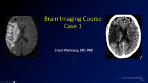
Case 1
83-year-old female with h/o hypertension presents with altered mental status, slurred speech, left hemiplegia and right sided gaze
Head CT/CTA
Top left – axial brain window
Top right – axial bone window
Bottom left – axial CTA
Brain MRI
Top row (left to right) – diffusion (DWI), ADC
Bottom row (left to right) – FLAIR, T1 postcontrast
Questions
1a. What study should be performed first in evaluating stroke?
1b. Which intracranial artery supplies the largest vascular territory of the major cerebral arteries?
Explanation video – Case 1
The video explanation for the brain course case 1 is here.
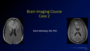
Case 2
60 year-old man with personality changes and lack of motivation with flat affect for 1-2 months
Brain MRI
Top row (left to right) – diffusion (DWI), FLAIR, T2, SWI
Bottom row (left to right) – sagittal T1 precontrast, axial T1 precontrast, axial T1 postcontrast, coronal T1 postcontrast
Questions
2a. What imaging characteristics in this case make this tumor appear higher grade?
Explanation video – Case 2
The video explanation for the brain course case 1 is here.
Case 3
62 year-old male with new onset of left sided weakness, left facial droop and right eye pain
Head CT
Top left – axial brain window
Top right – axial bone window
Bottom left – coronal brain window
Bottom right – sagittal brain window
Questions
3a. CTA is negative for aneurysm or arteriovenous malformation. What is your next step in management?
3b. In a patient with intraventricular extension of hemorrhage, what major complication are they likely to experience?
Case 4
65 year-old male with possible ground level fall found to be altered. On Coumadin for DVT.
Head CT
Top left – axial brain window
Top right – axial bone window
Bottom left – coronal brain window
Bottom right – sagittal brain window
Questions
4a. What additional complications do you see in this case?
4b. What territory is the infarct in?
Case 5
52 year-old with left sided weakness and numbness
Head CT
left – axial brain window
right – axial bone window
Brain MRI
Top row (left to right) – diffusion (DWI), FLAIR, T2
Bottom row (left to right) – T1 axial precontrast, T1 axial postcontrast, T1 coronal postcontrast
Questions
5a. Is this lesion intra-axial or extra-axial? How do you know?
Case 6
69 year-old-female with h/o autoimmune hepatitis admitted for hyponatremia. Hospital course complicated by vision changes.
Brain MRI
Top row (left to right) – diffusion (DWI), ADC, FLAIR
Bottom row (left to right) – T2 fat-saturated, T1 precontrast, T1 postcontrast
Questions
6a. How would you describe the restricted diffusion pattern and what is it concerning for?
Case 7
37 year-old-male presents with worst headache of life (WHOL)
Head CT/CTA
Top left – axial brain window
Top right – coronal brain window
Bottom left – axial CTA
Bottom right – coronal MIP CTA
Questions
7a. What is the cause of this hemorrhage?
7b. What is a well-known complication of subarachnoid hemorrhage (SAH)?When does it usually occur?
Summary
Hopefully by working through this course, you have learned:
- Modalities used in brain imaging
- Choosing an appropriate imaging modality
- Basics of reviewing brain cases
- Common imaging pathology of the brain
Remember, when considering imaging modalities
- CT – screening exam, good for finding major pathology
- MRI – more detailed for soft tissue of the brain parenchyma
Then, you were able to apply those principles in a case-based review. Now hopefully you can now recognize some of the key imaging findings of:
- Stroke
- Hemorrhage
- Tumor
- Infection
- Hydrocephalus
Thanks for joining us for this course, and please be sure to revisit the site and check out the YouTube Channel for additional learning resources.
If you haven’t already checked it out, there is a similar course for vascular imaging of the head and neck, so check it out as well.