Stroke vascular distributions – Imaging Case Review

Dr. Bailey is back for a case-based review of stroke and the vascular distributions commonly seen in stroke.

Introduction

In this video, we’ll review vascular territories in the brain as well as typical appearance of acute infarcts. This covers the distribution of the anterior cerebral artery (ACA), middle cerebral artery (MCA), posterior cerebral artery (PCA), cerebellar arteries, and basilar artery.

Case 1 – ACA infarct

This case shows an infarct in the right anterior cerebral artery distribution. There is loss of gray white differentiation on the CT. On MRI, it is even more apparent, with DWI abnormality which is dark on ADC. There is a corresponding abrupt occlusion of the ACA.

Case 2 – MCA infarct

In this case, there is hypoattenuation in the left posterior temporal lobe and inferior parietal lobe and posterior insular cortex. The MRI confirms that there is a stroke in this region. This is the posterior MCA distribution, with a posterior M2 branch occlusion

Case 3 – PCA infarct

There is subtle hypodensity in the left occipital lobe seen both on axial and sagittal CT. This is again confirmed on MRI, where there is T2 hyperintensity and diffusion abnormality. The MRA shows an abrupt cutoff of the left PCA.

Case 4 – Cerebellar infarct

This case shows a small, wedge shaped hypodensity in the left inferior cerebellum. MRI confirms abnormal diffusion in the left inferior cerebellum. In this case, the neck MRA shows

Case 5 – Multiple infarcts

This case shows multiple infarcts, including a right occipital and a left frontal infarct. When you have infarcts in multiple vascular territories, you should consider the possibility of a central source of thrombi, such as atrial fibrillation or cardiac disease, or vasculitis.

Case 6 – PCA plus

This case has an infarct in the left occipital lobe, but there is also hypoattenuation in the left midbrain and cerebral peduncle. MRI reveals even more areas of ischemia, including a small area in the right occipital lobe and multiple areas in the left thalamus. This indicates that the occlusion is more proximal and likely includes the basilar artery.

Case 7 – Medulla

This is a specific location which is frequently involved in infarcts, the lateral medulla. There is associated severe stenosis of the right vertebral artery.

Case 8 – Border zone

These are often seen as linear low attenuation along the border between vascular territories. In this case, it is the border between the ACA and MCA territories.

Special bonus case – artery of Percheron

This bonus case shows bilateral thalamic infarcts from an artery of percheron, a variant where the arterial supply for both thalami comes from a perforating branch on one PCA. This can also come from central venous thrombosis, so that is the other consideration

Special bonus case – venous infarct

If you have an infarction in an unusual location, particularly if associated with hemorrhage, then think about the possibility of sinus thrombosis. In this case, the straight sinus is dense and occluded on an MRV.

Summary

Hopefully these cases taught you something about the common locations of infarcts and their typical appearance on CT. Please check out the rest of the vascular and stroke content on the site.

See this and other videos on our Youtube channel

Brain Vascular Malformations

In this video, Dr. Bailey discusses the most common vascular malformations and reviews the most common grading system for arteriovenous malformations (AVMs), the Spetzler-Martin grading scale.

Introduction to arteriovenous malformations

Arteriovenous malformations are vascular anomalies consisting of feeding arteries, a nidus where the shunt is located, and one or more draining veins. AVMs can be compact or have a diffuse nidus. There can be surrounding gliosis and potentially calcification on CT or calcium sensitive imaging. Imaging will demonstrate flow voids,

Spetzler-Martin grading scale

The Spetzler-Martin scale gives a score between 1-5, with points assigned based on size (< 3 cm, 1 point; 3-6 cm, 2 points, and > 6 cm, 3 points), involvement of eloquent cortex (1 point), and involvement of deep veins (1 point). This score can help predict the potential surgical morbidity and mortality.

Arteriovenous fistulas

Arteriovenous fistulas (AVFs) are abnormal shunts most commonly from dural vessels. These are abnormal connections between these arteries and the dural venous drainage. Often external carotid artery branches will be dilated as they are the abnormal supply. There is arterialization of the dural venous sinuses. These are most common at the transverse-sigmoid sinus junction.

Cavernous malformations

Cavernous malformations are slow flow venous malformations that have well contained abnormal veins and vessels. They have areas of hemosiderin with T1 hyperintensity, T2 hyperintensity centrally and a peripheral hemosiderin rim. They may have an abnormal adjacent vessel or developmental venous anomaly (DVA). On CT, they may be hyperdense and can be confused with hemorrhage, but central calcification is a good clue. Multiple cavernous malformations can occur in familial syndromes.

Developmental venous anomaly (DVA)

DVAs are congenital venous malformations draining normal veins. These are the most common vascular malformation and are benign. They appear as a branching tree of abnormal venous drainage going to normal veins.

Capillary telangiectasia

These are slow flow capillary malformations that are incidentally found. They have stippled enhancement and you may see something on the blood sensitive imaging (GRE or SWI). There is usually no abnormal edema or FLAIR.

Thanks for tuning in to this video about intracranial vascular malformations. Please check out the additional vascular videos on the site.

See this and other videos on our Youtube channel
Circle of Willis

In this video, Dr. Bailey reviews the anatomy of the Circle of Willis, or the confluence of the internal cerebral and basilar arteries within the brain. She reviews the normal anatomy, talks about some common variants you may encounter, and introduces a few less common variants.

Introduction to the Circle of Willis

The Circle of Willis is the circular anatomical construct of vessels made up by the internal carotid arteries, the basilar artery, and their intracranial proximal branches. This includes the anterior and posterior communicating arteries and the anterior, middle, and posterior cerebral arteries.

Posterior circulation

The posterior circulation includes the posterior cerebral arteries, the basilar artery, the superior cerebellar artery, the anterior inferior cerebellar artery (AICA), and the posterior inferior cerebellar artery (PICA). The AICA is particularly variable and may be hard to see, particularly on 3D imaging. The PICA arises from the vertebral artery and may also vary in size from one side to the other.

Common variants

One particularly common variant is a vertebral artery that terminates in PICA. That is, there is either no or a very vertebral artery is seen distal to the PICA origin. Dolichoectasia is a tortuous and prominent basilar artery larger than 4.5 mm in transverse diameter. It is also a common variant to have no posterior communicating arteries (P-comms). A fetal PCA, is vessel that arises from the posterior communicating artery with an absent or very small P1 segment of the PCA. A hypoplastic A1 is a small A1 on one side, with both A2 segments arising from one side. The A1 segments may arise at various levels and be tortuous. An azygous ACA is a single, or unpaired, ACA in the A2 segment where both sides fuse and there is a common ACA. Sometimes you can have the opposite and have 3 A2 segments. Any of the arteries can also be duplicated, or you can have a fenestration, a small wall within the center of the vessel. Fenestrations can mimic thrombus but they are often very linear along the course of the vessel.

Less common variants

The persistent trigeminal artery is a persistent fetal connection between the anterior and posterior circulation at the level of trigeminal artery. It is the most common persistent fetal connection and passes through Meckel’s cave (the trigeminal cistern).  

It’s also possible to see a missing vessel, such as an absent ICA. In these cases, they may be congenitally absent or chronically occluded.

Thanks for tuning in to this video about the normal and variant anatomy of the circle of willis. Please check out the vascular imaging page on the site.

See this and other videos on our Youtube channel.

Intracranial aneurysms

In this video, Dr. Bailey reviews intracranial aneurysms, including an overview of what an aneurysm is, how to find them, and tips for searching for aneurysms. The first part of this video covers general principles of aneurysm evaluation and the second part shows 3 sample cases that you can go through to test your individual skills.

Introduction and definition of aneurysm

An aneurysm is an abnormal outpouching of the intracranial vessel. Intracranial aneurysms are true aneurysms, which typically involve all of the layers of the vessel. To find aneurysms, a type of vascular imaging known as an angiogram (which can also be done with CT or MRI) is done to evaluate the arteries.

How to find aneurysms

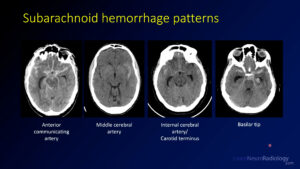
To find aneurysms, you should be looking for an abnormal dilation of the vessel. Then you should look at the most common locations, which are the anterior communicating artery, the carotid terminus/posterior communicating artery, the middle cerebral artery, and the posterior circulation/basilar tip.

Saccular versus fusiform aneurysm

Saccular aneurysms are the most common type of aneurysm. They are rounded outpouchings of the vessel. Fusiform aneurysms are circumferential areas of enlargement of the vessel, meaning the whole diameter is increased.

Thrombosed aneurysm

Sometimes aneurysms can be thrombosed, meaning they no longer have blood flow. In these cases, their imaging appearance can change, and they may not show up on angiographic imaging.

Tips for reporting aneurysms

When creating a report about an aneurysm, you should describe where the aneurysm is, which direction it points, and its size. It’s common to measure three sizes, the maximal width, the height, and the width at the neck.

Aneurysm treatment

The two most common treatments for aneurysm are surgical clipping, in which a clamp is placed over the base of the aneurysm, and endovascular coiling, in which small coils are placed within the vessel from inside. It is also possible to use stents to treat aneurysms, either in combination with coils or alone.

Practice cases

Take a crack at 3 practice cases at the end of the video.

Hopefully you learned a little bit about finding and reporting intracranial aneurysms. Please check out the additional vascular videos on the site, including the video on 5 quick ways to improve your aneurysm search pattern.

See this and other videos on our Youtube channel
Vascular Imaging – Playlist

This playlist covers a range of topics in vascular imaging including general concepts about how to approach brain and neck vascular imaging, what intracranial aneurysms, are, and how to improve your aneurysm search pattern.

You can learn more about other concepts in vessel imaging and other abnormalities on the vascular imaging page. If you haven’t already, you might consider taking a look at the vascular imaging capstone course. Also, please check out our full channel on Youtube.

5 ways to improve your brain aneurysm search pattern

In this video, I walk you through 5 quick tips that you might use to improve your brain aneurysm search pattern on CT angiograms of the brain. This is a longer version of a lecture I put together with Everlight Radiology, so be sure to check them out.

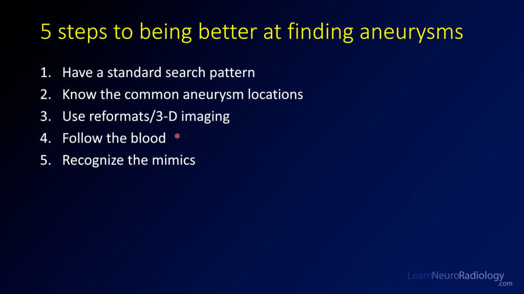
Have a standard search pattern. When I’m looking at a CTA of the head, I do the anterior circulation first and then move from right to left, then over to the posterior circulation.

Know the common aneurysm locations. The most common aneurysm location is the anterior communicating artery (35%) followed by the carotid terminus (30%) and middle cerebral artery (20%). Posterior circulation aneurysms are relatively uncommon (10%) but it’s important to look there as well. Try to use these tips on the sample case.

Use reformats and 3-D imaging. These supplemental tools can help you improve your sensitivity. Multiplanar reformats are thin slices that are displayed in the other planes, while maximum intensity projections (or MIPs) show you the brightest pixel in a thicker slice. Volume renderings are a nice way to make measurements and increase your sensitivity.

Using the MIPs can definitely make you more sensitive. The axial MIPS are great to see the MCAs, the sagittal MIPs are great to see the carotid terminus and ACAs, and the coronal MIPs are great to see the posterior circulation and MCAs again.

Follow the blood. This is my favorite tip. The location of the blood on the non-contrast CT is one of the best clues about where your aneurysm is going to be. You need to check that area very closely.


Recognize the mimics. There are some things which can mimic aneurysmal subarachnoid hemorrhage, but some features may help you know that it is less likely to be from an aneurysm. Atypical location, an unusual history, or unusual patient demographics can clue you in that it might be a different cause. Be sure to think about hypertensive hemorrhage, venous infarct, tumor (glioma, metastasis, or cavernous malformation), and benign perimesencephalic hemorrhage.

Summary. These 5 quick tips can help you be better at understanding aneurysms and being better and finding them.

If you haven’t already, be sure to check out the vascular imaging course and sample cases that you can scroll through.

See all of the search pattern videos on the Search Pattern Playlist.

Vascular Imaging of the Head and Neck – Case D

his case is the fourth and final case that goes with the vascular capstone course. On that page, there is a scrollable case that you can go through to teach you how to approach a CTA of the head in a real patient.

This case is a 41 year-old man after a trauma in a motor vehicle collision (MVC). Take a look and see what you think before continuing on (https://bit.ly/CTAcaseD).

Starting with the CTA of the neck, this is not a normal case. If you follow your normal search pattern, you will see that there are a number of abnormalities, starting from the right internal carotid artery (ICA), which is lumpy and irregular looking. The left ICA is worse, with areas of narrowing, outpouchings, and linear filling defects that represent little areas of the intima that are lifted up by trauma. The little outpouchings along the margins of the vessel are little pseudoaneurysms, or areas where the vessel is injured and contrast is able to leak out into the surrounding area of damaged vessel. Both vertebral arteries are also abnormal with multifocal irregularity and a small pseudoaneurysm on the right.

This is a dramatic example of traumatic vascular injury in the neck. After high energy or penetrating (think gunshot or stab wound), the great vessels can be injured and jeopardize the blood supply to the brain. These injuries are graded on the Denver, or Biffl, scale which ranges from 1-5. You can read more about it on Radiopaedia. Injuries to the vessels in the neck are most commonly pseudoaneurysms, meaning that one layer or more of the vessel is injured and the wall of the aneurysm does not contain all the layers (intima-media-adventitia). Contrast this to intracranial aneurysms, which are true aneurysms and the wall contains all the layers.

This is the last of the case examples for the vascular capstone course. If you haven’t already, I recommend going back to the vascular capstone course, where you can review the other browseable cases with explanations.

The capstone overview is here, if you’d like to see all the cases and videos.

Or, see all of the vascular capstone videos in the vascular imaging capstone playlist.

Vascular Imaging of the Head and Neck – Case C

This case is the third of four cases that go with the vascular capstone course. On that page, there is a scrollable case that you can go through to teach you how to approach a CTA of the head in a real patient.

This case is a 43 year-old woman with seizure. Take a look and see what you think before continuing on ( https://bit.ly/CTAcaseC).

On the noncontrast CT, you see some high density material in the right frontal lobe. It is more dense than contrast, which is consistent with calcium. If you look more closely at the morphology, it is almost curvilinear or tubular, giving the impression that it might be calcification along vascular structures. The sulci of the frontal lobe are effaced with masslike effect on the brain.

While the diagnosis is uncertain, there are enough features to make us think that there is an underlying vascular malformation. To check, we need to do some sort of vascular imaging. The most readily available and fastest is to do a CT angiogram.

On the CT angiogram, the proximal vessels are normal, but the abnormalities start at the proximal middle cerebral artery (MCA) and anterior cerebral artery (ACA) on the right. There are too many vessels in this region and they are too tortuous. If you follow them up, they go to a large and tortuous vascular abnormality in the right frontal lobe.There are enlarged draining veins that empty into the superior sagittal sinus.

This is the classic imaging appearance of arteriovenous malformation (AVM) within the brain. AVMs are congenital vascular abnormalities that consist of an abnormal connection of arteries and veins with a tangle of abnormal vessels, the nidus. Go on and look at the volume rendered (VR) reconstruction and the coronal maximum intensity projection (MIP) images to better understand what this abnormality looks like.

Once you’ve finished this video, I recommend going back to the vascular capstone course, where you can review the other browseable cases with explanations. The capstone overview is here , if you’d like to see all the cases and videos.

Or, see all of the vascular capstone videos in the vascular imaging capstone playlist.

Vascular Imaging of the Head and Neck – Case B

This case is the second of four cases that go with the vascular capstone course. On that page, there is a scrollable case that you can go through to teach you how to approach a CTA of the head in a real patient.

This case is a 47 year-old woman with new neurologic symptoms. Take a look and see what you think before continuing on (https://bit.ly/CTAcaseB).

The patient in this case has scattered small infarcts in the left MCA distribution on diffusion weighted imaging from an MRI (top left window). This raises suspicion for an underlying vascular abnormality. Remember, for stroke and vascular supply issues, you want to include CTA of the neck because these vessels supply the vessels of the circle of Willis.

On the CTA, you see multiple abnormalities. The right internal carotid artery (ICA) is tortuous and irregular in the superior neck, which is highly atypical for a patient of this age. The left internal carotid artery (ICA) is even more abnormal, with smooth tapering of the vessel to severe narrowing just above the bifurcation. The low density filling defect is thrombus, some of which is probably under a dissection flap and some of which is free-floating in the vessel. The more distal ICA also has tortuosity and narrowing similar to the right, confirming the abnormality is bilateral.

Don’t forget to complete your search pattern, but the vertebral arteries and intracranial vessels are pretty normal.

Given the multiple areas of vascular narrowing of the cervical carotid in a middle age woman with an ICA dissection on the left, this patient most likely has fibromuscular dysplasia (FMD). Fibromuscular dysplasia is the most common vasculopathy in middle-aged women and frequently affects the renal arteries, ICAs, and vertebral arteries. Characteristic findings are the beads on a string appearance of the vessel with multifocal areas of narrowing interspersed with dilation.

Once you’ve finished this video, I recommend going back to the vascular capstone course, where you can review the other browseable cases with explanations. The capstone overview is here, if you’d like to see all the cases and videos.

Or, see all of the vascular capstone videos in the vascular imaging capstone playlist.

Vascular Imaging of the Head and Neck – Case A

This case is the first of four cases that go with the vascular capstone course. On that page, there is a scrollable case that you can go through to teach you how to approach a CTA of the head in a real patient.

This case is an 80 year-old woman who presented with altered mental status. Take a look and see what you think before continuing on.

The patient in this case has subarachnoid and parenchymal hemorrhage on a noncontrast CT. Because aneurysms and vascular malformations are possible causes of subarachnoid hemorrhage, we proceeded with vascular imaging, or a CT angiogram of the head, to look for aneurysms or other possible vascular causes. Remember, for an intracranial hemorrhage you don’t need the CTA of the neck because these don’t commonly have any pathology that can explain intracranial hemorrhage.

On the CTA, you see multiple abnormal outpouchings of the intracranial vessels, otherwise known as an aneurysm. Intracranial aneurysms are abnormal outpouchings of the vessels thatt contain all the layers of the vessel wall (true aneurysms). They have a risk of rupture of several percent per year, and can be treated with surgical clipping or endovascular methods such as coils. Remember, it is common for patients to have more than one aneurysm, as is seen in this case.

Once you’ve finished this video, I recommend going back to the vascular capstone course, where you can review the other browseable cases with explanations. The capstone overview is here, if you’d like to see all the cases and videos.

Or, see all of the vascular capstone videos in the vascular imaging capstone playlist.