Emergency Imaging of Brain Tumors
You can learn more about brain tumors on the brain tumor topic page. Also, please check out our full channel on Youtube.
Author: Brent Weinberg, MD
You can learn more about brain tumors on the brain tumor topic page. Also, please check out our full channel on Youtube.
In this video, I walk you through 5 quick tips that you might use to improve your brain aneurysm search pattern on CT angiograms of the brain. This is a longer version of a lecture I put together with Everlight Radiology, so be sure to check them out.
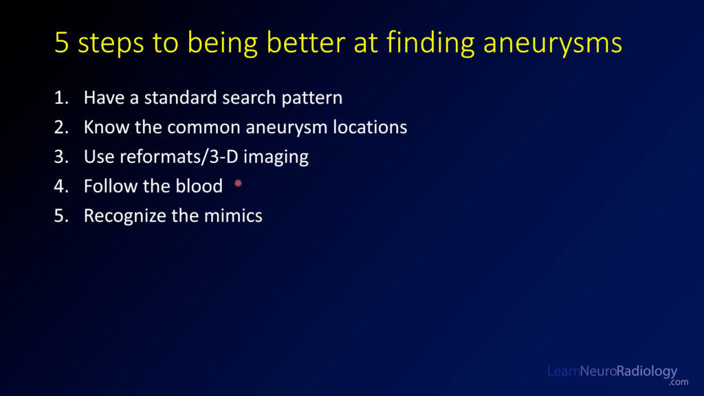
Have a standard search pattern. When I’m looking at a CTA of the head, I do the anterior circulation first and then move from right to left, then over to the posterior circulation.
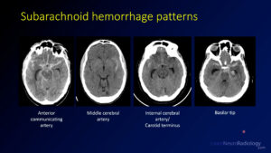
Know the common aneurysm locations. The most common aneurysm location is the anterior communicating artery (35%) followed by the carotid terminus (30%) and middle cerebral artery (20%). Posterior circulation aneurysms are relatively uncommon (10%) but it’s important to look there as well. Try to use these tips on the sample case.
Use reformats and 3-D imaging. These supplemental tools can help you improve your sensitivity. Multiplanar reformats are thin slices that are displayed in the other planes, while maximum intensity projections (or MIPs) show you the brightest pixel in a thicker slice. Volume renderings are a nice way to make measurements and increase your sensitivity.
Using the MIPs can definitely make you more sensitive. The axial MIPS are great to see the MCAs, the sagittal MIPs are great to see the carotid terminus and ACAs, and the coronal MIPs are great to see the posterior circulation and MCAs again.
Follow the blood. This is my favorite tip. The location of the blood on the non-contrast CT is one of the best clues about where your aneurysm is going to be. You need to check that area very closely.

Recognize the mimics. There are some things which can mimic aneurysmal subarachnoid hemorrhage, but some features may help you know that it is less likely to be from an aneurysm. Atypical location, an unusual history, or unusual patient demographics can clue you in that it might be a different cause. Be sure to think about hypertensive hemorrhage, venous infarct, tumor (glioma, metastasis, or cavernous malformation), and benign perimesencephalic hemorrhage.
Summary. These 5 quick tips can help you be better at understanding aneurysms and being better and finding them.

If you haven’t already, be sure to check out the vascular imaging course and sample cases that you can scroll through.
See all of the search pattern videos on the Search Pattern Playlist.
Wondering where all the junk on the internet comes from? Apparently it is written by computers using artificial intelligence! Well, at least that’s what the folks over at jasper.ai would like for you to believe. According to this company, they are using AI to help you write materials for your blog or website to drive traffic your direction. At the very least, they promise they will make it easier for you.
People have been promising that computers were going to take over radiology for at least the past decade, and as far as I can tell there has been very little progress. However, most of this is about image interpretation and this is the first time I’ve seen a product claim that it can do writing for you.
As the owner of learnneuroradiology.com and producer of a lot of educational content, I was wondering what this would mean for someone who creates highly specialized content like myself. I figured this might be halfway decent for a generic interest blog or website, but I didn’t think it would be very good for subspecialized material like radiology and specifically neuroradiology.
I started by taking a look at their introduction video, where they make a lot of claims about how much faster they can create content and show you a brief example. Like all promotional material, it definitely makes big promises, including that they have analyzed 10% of the internet. There are testimonials and everything. I feel like this tells us a lot about the internet that most of it is being written by a bot.
It took a little bit to set up a trial. I had to enter the name of my website, some billing information (including a credit card number), and what kind of content I was creating. The full product starts at $49 / month but there is a free trial for 5 days. That’s what I’m taking. Once I finished, I was able to see the full dashboard.
I started with a generic article about Brain MRI to see how it would do with some more general content. After entering some basic information, I got started pretty quickly. It required me to start writing the article before it created some content, but surprisingly it generated some half-way relevant, if overly generic material. With a little bit of guidance and a few clicks, I had created a decent general interest article. My initial impressions were that it was doing ok. It did a pretty decent job on a general article. I give it a “B to B+”.
Now it’s time to give it something a little harder. Glioblastoma. I expected it to perform worse, but it had some surprisingly decent comments about the imaging features and could even differentiate between imaging modalities, like computed tomography, magnetic resonance imaging. It could fill in rudimentary although sometimes wrong information about the differential diagnosis and prognosis. I’m not going to lie, this is exceeding my expectations.
Finally, I tried an article a little bit more technical about white matter abnormalities. Of all the articles, this one did the worst, but it was still fairly relevant. It was able to come up with some differential diagnosis for white matter lesions and relevant diseases. It did provide a little bit more irrelevant or wrong content than the other articles.
It did make me a little sad that we don’t have more report generation tools that are radiology specific. I feel like a similar tool trained on radiology reports with radiology diagnoses could actually go a long way towards helping me generate differential diagnoses on challenging cases. I’m looking forward to having more tools like this in the future but I don’t feel like this is quite ready for primetime right now, even for writing a website.
Tune in next time for additional interesting content and radiology teaching material! Thanks for checking out the site!
So what are my final recommendations? I wouldn’t use it for my site, but it is capable of generating some half-way useful content. I expected to be able to make fun of it more, but it exceeded my applications. What is my overall impression: I wouldn’t throw it in the trash. I can imagine it is pretty useful for a generic interest site, but for more specialized applications it does get a little unraveled. It becomes a little repetitive, and I’m worried that it is actually just regurgitating content from other sites. There is a “plagiarism checker” but I was unable to use it because it required an additional fee that I didn’t want to pay.
“What is my overall impression: I wouldn’t throw it in the trash.”
It did make me a little sad that we don’t have more report generation tools that are radiology specific. I feel like a similar tool trained on radiology reports with radiology diagnoses could actually go a long way towards helping me generate differential diagnoses on challenging cases. I’m looking forward to having more tools like this in the future but I don’t feel like this is quite ready for primetime right now, even for writing a website.
Tune in next time for additional interesting content and radiology teaching material! Thanks for checking out the site!
Neuroradiology brain tumor board review. This lecture is geared towards the ABR core exam for residents, but it would be useful for review for the ABR certifying exam or certificate of added qualification (CAQ) exam for neuroradiology.
This video has both the final case of the series and a quick review summary!
In this case, you are starting with an immunocompromised patient with HIV. Initial CT images show a hyperdense mass in the left basal ganglia with a lot of surrounding edema. This is helpful, because a few things are known for being hyperdense on CT.
MRI images confirm a mass in the basal ganglia. It is somewhat T2 hypointense with well-defined margins and surrounding edema. On postcontrast images, it has peripheral enhancement but central non-enhancement compatible with necrosis.
The differential diagnosis for a solitary enhancing parenchymal mass is different in an immunocompromised patient (or someone on immune suppressing agents. In an immune normal patient, the top diagnoses are
On the other hand in an immunocompromised patient the order of these diagnoses shifts to include:
As you can see, lymphoma and infection jump to the top in an immunocompromised patient.
The diagnosis is: CNS lymphoma
CNS lymphoma can occur when associated with systemic lymphoma or primarily in the CNS, as in this case. This is most commonly a diffuse large B-cell lymphoma. It is more common in immune compromised patients. It often occurs in the basal ganglia and periventricular white matter and can often be multifocal. Lymphoma is one of the rare diseases which is T2 hypointense, so you should think about it if you see a T2 hypointense mass.
In immunocompetent patients, lymphoma most commonly has solid enhancement. However, in immunocompromised patients it is much more likely to show central necrosis, as in this case. Also, in an immunocompromised patient, it can be hard to differentiate lymphoma from infection, particularly toxoplasmosis. The two most common ways to try to differentiate this are to start a trial of toxoplasmosis therapy for a few weeks and see if the lesions improve and to perform a thallium-201 chloride nuclear medicine scan. Lymphoma has thallium uptake, while toxoplasmosis does not.
In this board review lecture, you’ve seen a lot of different tumors and how they manifest in different situations. In many cases, you can’t make a definitive diagnosis but you should always be able to come up with a reasonable differential diagnosis. It’s also helpful to know some of the basics about treatment and prognostic factors.
There are two key strategies that I hope can help you get a few additional points, the approach to CP angle masses and the approach to cortical tumors.
As we’ve seen in some of the other cases, cerebellopontine angle masses can be solid or cystic. Solid masses that involve the IAC and expand it are likely schwannomas, while others outside the IAC are likely meningiomas. Arachnoid cysts and epidermoids are the most common cystic masses which are differentiated by DWI (which is bright in ependymomas.
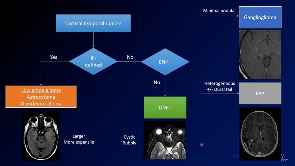
Several of the cases in this series dealt with cortical temporal tumors. Ill-defined masses that are larger are more likely to be low grade gliomas (oligodendrogliomas and astrocytomas). Completely non-enhancing bubbly masses favor DNET. A little nodular enhancement favors ganglioglioma, while pleomorphic xanthoastrocytoma (PXA) can be more avidly enhancing and irregular.
Neuroradiology brain tumor board review. This lecture is geared towards the ABR core exam for residents, but it would be useful for review for the ABR certifying exam or certificate of added qualification (CAQ) exam for neuroradiology.
More description and the answer (spoiler!) are seen below the video.
This case of a patient with diplopia (double vision) starts with a CT through the orbits. You can see there is a well-defined mass at the right orbital apex.
On MRI, you can see the lesion again is well-defined with well defined margins and is hyperintense on T2. On T1, the lesion is isointense to muscle on pre-contrast and then demonstrates heterogeneous enhancement on postcontrast. It looks like the lesion is enhancing more on the coronal image compared to the axial image.
The diagnosis is: orbital venous vascular malformation
Orbital venous vascular malformations are sometimes referred to as hemangiomas, although this term is falling out of favor because it is not neoplastic (unlike the neoplastic infantile and neonatal orbital hemangiomas). These are relatively benign lesions that can cause visual problems secondary to mass effect, but it’s relatively uncommon for them to enlarge.
One characteristic finding of orbital venous malformations is progressive enhancement on delayed images. On early images, it might be enhancing a little bit but if you have more delayed images, you might see more enhancement. If you don’t have more than one plane of contrast, you can bring them back and image them again 15-30 minutes later and it should fill in.
The primary differential considerations for orbital masses are sarcoidosis, idiopathic or IgG4 related orbital disease, metastatic disease, and lymphoma. When it is this well defined and has the characteristic delayed enhancement, the diagnosis is relatively certain.
Neuroradiology brain tumor board review. This lecture is geared towards the ABR core exam for residents, but it would be useful for review for the ABR certifying exam or certificate of added qualification (CAQ) exam for neuroradiology.
More description and the answer (spoiler!) are seen below the video.
This case starts with 2 axial images from a CT through the posterior fossa followed by MRI images through the same region. There is a heterogeneous lesion to the left of the midline along the left foramen of Lushka. On postcontrast images, it is pretty avidly enhancing. The enhancing margins are pretty well defined and it looks like it is wholly in the ventricle.
The diagnosis is: ependymoma
Ependymomas are enhancing intraventricular tumors arising from the ependymal lining. They are commonly enhancing and conform to the ventricles and the ventricular outflow tract, which results in their description of “toothpaste” like lesions. In adults, they most commonly occur in the 4th ventricle although in pediatric patients they can occur elsewhere.
When in the posterior fossa, your main differential is choroid plexus papilloma/tumor. If you can’t tell that it’s an intraventricular lesion it can be harder because the differential diagnosis also include metastatic disease and possibly medulloblastoma. If you see a lesion that looks similar but doesn’t enhance very much, think about it’s sister lesion subependymoma.
Neuroradiology brain tumor board review. This lecture is geared towards the ABR core exam for residents, but it would be useful for review for the ABR certifying exam or certificate of added qualification (CAQ) exam for neuroradiology.
More description and the answer (spoiler!) are seen below the video.
In this case, we have an MRI showing a FLAIR and T2 hyperintense mass in the left insula with relatively ill-defined margins. On SWI, there are some areas of susceptibility that probably represent calcification, although blood products could look similar. Postcontrast images demonstrate little or no contrast enhancement.
1:33 The diagnosis is: oligodendroglioma
Oligodendrogliomas are gliomas which are now defined by the characteristic genetic features of IDH mutation and 1p19q codeletion (loss of portions of both chromosomes 1 and 19). They can be WHO grade 2 (as in this case) or grade 3 (anaplastic oligodendroglioma). Theoretically, these lesions never degrade into WHO grade 4 lesions although the grade 3 lesions can be quite aggressive. In general, oligodendrogliomas have a better prognosis than their sister gliomas, astrocytomas. They respond better to radiation and have better overall survival.
Oligodendrogliomas are treated with a combination of resection and chemoradiotherapy.
The susceptibility seen within the tumor on this case represents areas of calcification. Oligodendrogliomas are one of the main considerations if you see an expansile tumor with calcification.
Neuroradiology brain tumor board review. This lecture is geared towards the ABR core exam for residents, but it would be useful for review for the ABR certifying exam or certificate of added qualification (CAQ) exam for neuroradiology.
More description and the answer (spoiler!) are seen below the video.
This case shows a 20 year-old with seizures. MRI shows a lesion in the medial temporal lobe along the tentorium. It is relatively well circumscribed with a cystic portion as well as an enhancing nodule. There is not much mass effect.
The diagnosis is: ganglioglioma
Gangliogliomas are low grade tumors often found in the temporal lobes of patients with seizures. They are mixed in their cell origin, containing both components of glial and neuronal cells. Along with pilocytic astroctyoma, PXA, and hemangioblastoma, they are one of the tumors in the differential for a tumor with a cyst and a nodule. They can be impossible to differentiate from DNET, but if you see enhancement it is more likely to be a ganglioglioma. Gangliogliomas are the most common neoplastic cause of epilepsy.
When you see a minimally enhancing cortical tumor, you should have a relatively short differential which includes:
First consider whether they are ill-defined or well-marginated. If ill-defined, the differential includes astrocytoma or oligodendroglioma. If well-marginated, then consider whether there is enhancement. If no enhancement, DNET is most likely. If there is a small amount of nodular enhancement, favor ganglioglioma, as in this case.

In a testing situation, if a small and minimally enhancing cortical tumor enhances a little bit, choose ganglioglioma. If you don’t seen enhancement, choose DNET. PXAs tend to be much more heterogeneous and irregular.
If you use a structured approach to these tumors, you can fall back on it if you aren’t really sure what you are looking at. With these relatively simple rules, you can be sure to get the most points on your exams AND give the most meaningful differential diagnosis.
Neuroradiology brain tumor board review. This lecture is geared towards the ABR core exam for residents, but it would be useful for review for the ABR certifying exam or certificate of added qualification (CAQ) exam for neuroradiology.
More description and the answer (spoiler!) are seen below the video.
As we start this case, we see a brain and a bone window from a CT through the posterior fossa. It is a little bit hard to see because it is a midline abnormality. The sagittal and coronal reformats are helpful in identifying where the lesion is located.
MRI is a little bit easier to see the lesion, but only a little. There is a well-demarcated lesion along the inferior aspect of the 4th ventricle. There is calfication, which you could see both on CT and GRE sequences from MRI. On post-contrast imaging, there is little, if any enhancement.
The diagnosis is: subependymoma
Subependymomas are relatively benign tumors arising from the walls of the ventricles. In contrast to most other intraventricular tumors, they often do not have much enhancement. The most common locations are in the 4th ventricle and lateral ventricles. Many times they are incidental but they can cause symptoms related to hydrocephalus.
Your differential diagnosis for intraventricular lesions includes meningioma, ependymoma (more enhancing), choroid plexus tumors (also more enhancing), and other tumors such as subependymal giant cell tumors (SEGT, often seen in patients with tuberous sclerosi).
Some tumor types have characteristic histologies that you should be familiar with for the test. Ependymomas are associated with perivascular pseudorosettes, while other tumors such as glioblastoma have their own histologic keywords (pseudopallisading necrosis, microvascular proliferation). These are pretty low yield to study but if you see them it’s good to be familiar with them.
Neuroradiology brain tumor board review. This lecture is geared towards the ABR core exam for residents, but it would be useful for review for the ABR certifying exam or certificate of added qualification (CAQ) exam for neuroradiology.
More description and the answer (spoiler!) are seen below the video.
This case shows an MRI with a mass in the left parietal region near the midline. It is pretty well circumscribed and is probably extra-axial. It is homogeneously enhancing on postcontrast, and both T2 and T1 images demonstrate significant vascular flow voids suggesting that this is a very vascular lesion.
The diagnosis is: solitary fibrous tumor
Solitary fibrous tumors of the CNS (previously known as hemangiopericytomas) are similar in histology to fibrous tumors elsewhere in the body, such as the pleura. These were originally thought to be related to meningiomas, but their genetics has proven this to be false. However, the imaging features are similar to aggressive meningiomas. They are most often extra-axial, have avid enhancement, and broad dural attachments. It is common to have flow voids showing a high degree of vascularity.
Like meningiomas, these can be highly vascular lesions with much of their blood supply coming from external carotid branches. This angiogram shows the high degree of vascularity. It can be favorable to embolize as many of these vessels as possible prior to surgical resection to minimize bleeding.
Remember that the nomenclature has changed but you still may run into the term hemangiopericytoma. It’s something that should be on your differential if you see something that looks like an aggressive meningioma.